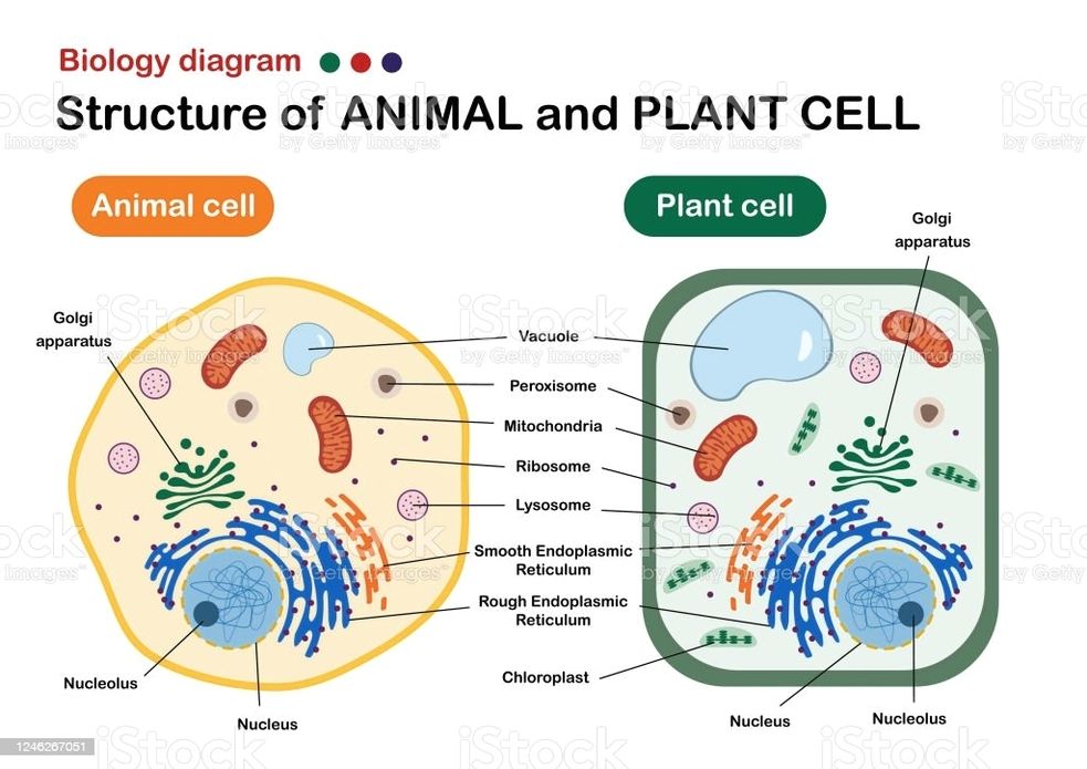
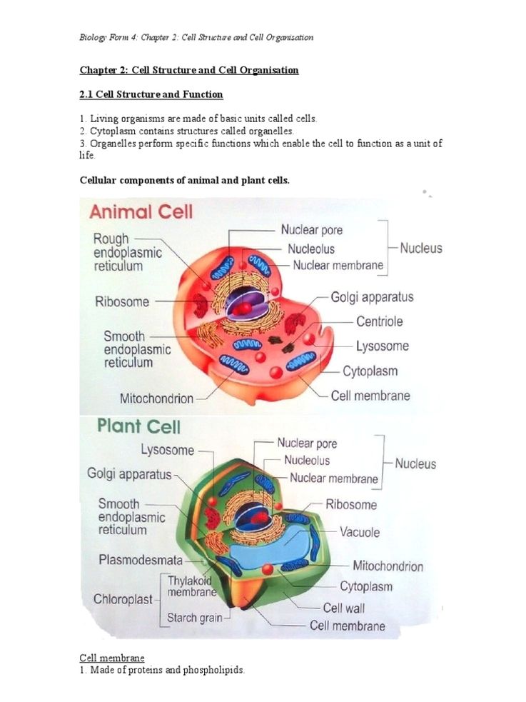
See the below image for the Biology show structure of animal and plant cell diagram. Plant and Animal Cell Structures 1 They are single membrane-bound, fluid-filled structures. 2 They are found in both plants and animal cells. Plant cells have large, central vacuole. … 3 Tonoplast is the membrane of these vacuoles. 4 They maintain turgor pressure.
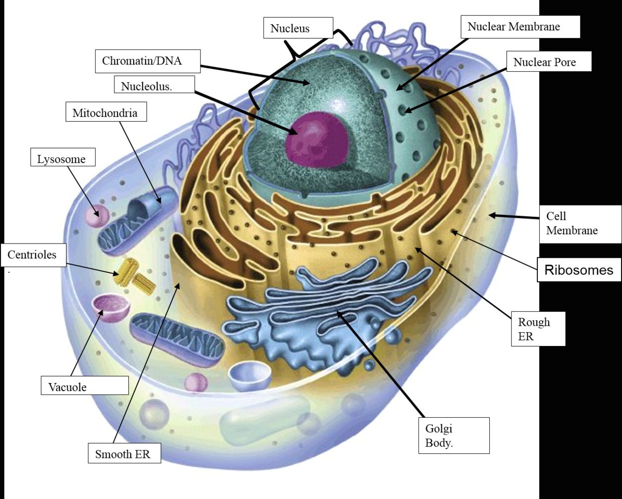
Structure of Animal and Plant Cells 1 Have a nucleus 1. Plant cells have a cellulose cell wall 2 Have a cytoplasm 2. Plant cells have a vacuole containing cell sap 3 Have a cell membrane 3. Plant cells have chloroplast 4 Contain mitochondria 4. Many plant cells have a box-like shape whilst animal cell shape varies 5 Contain ribosomes
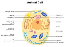
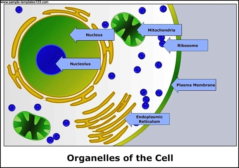
Generalized Structure of a Plant Cell Diagram. As you can see in the above labeled plant cell diagram under light microscope, there are 13 parts namely, Cell membrane. Cytoplasm. Ribosomes. Nucleus.