Functional Divisions Of Nervous System

Functional Divisions Of Nervous System: The nervous system is divided into the central nervous system (CNS), comprising the brain and spinal cord, and the peripheral nervous system (PNS), which includes all the nerves outside the CNS. The PNS is further divided into the somatic and autonomic nervous systems.

Functional Divisions Of Nervous System

Human Cell with Detailed Labels

Human Cell with Detailed Labels: A detailed diagram of a human cell includes organelles like the nucleus, nucleolus, ribosomes, mitochondria, endoplasmic reticulum, Golgi apparatus, lysosomes, peroxisomes, and centrosomes, along with the plasma membrane and cytoskeleton. Each component is labeled to reflect its function, such as energy production, protein synthesis, or genetic storage. These labels and structures offer insight into how cells operate as the fundamental units of life.

Human C

Multiple Cell Diagram with Labels

Multiple Cell Diagram with Labels: A diagram showing multiple cell types with labels often includes a variety such as nerve cells, muscle cells, blood cells, and epithelial cells, each with unique shapes and structures suited to their function. Nerve cells have long axons for signal transmission, muscle cells have fibrous structures for contraction, red blood cells are biconcave for efficient gas transport, and epithelial cells line surfaces for protection and secretion. This comparative diagram helps learners understand the diversity and specialization in multicellular organisms.

Biology C

Open and Closed Stomatal Pore

Open and Closed Stomatal Pore: Stomatal pores on leaf surfaces regulate gas exchange and water loss in plants. When open, stomata allow the intake of carbon dioxide necessary for photosynthesis and the release of oxygen and water vapor through transpiration. In closed states, which occur during water stress or high temperatures, the pores help conserve water by preventing evaporation. Guard cells on either side of the pore change shape to open or close the stomata in response to environmental cues. Diagrams depict both states, highlighting the dynamic role of stomata in plant physiology.

Biological Diagrams Description

Flower Anatomy Diagram

Flower Anatomy Diagram: A detailed flower anatomy diagram showcases both reproductive and supportive parts. The petals are brightly colored to attract pollinators, while the sepals protect the bud before it opens. The male reproductive part, the stamen, includes the anther, which produces pollen, and the filament, which supports it. The female part, the carpel (or pistil), consists of the stigma (where pollen lands), the style, and the ovary, which contains ovules. These labeled parts help explain plant reproduction and the pollination process, demonstrating the intricate design of flowers in botany.

Biological Diagrams Described

Animal Cell Diagram

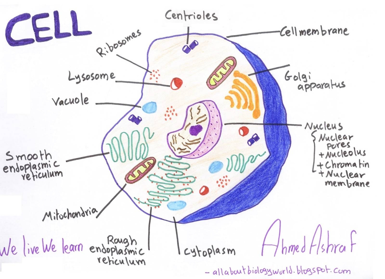
Animal Cell Diagram: An animal cell is a eukaryotic structure that contains several key organelles, each with specific functions crucial for the cell’s survival. The nucleus is the control center housing DNA, surrounded by the nuclear envelope. The cytoplasm is the gel-like fluid where all cellular components reside. The endoplasmic reticulum (smooth and rough) aids in the synthesis of lipids and proteins. The Golgi apparatus processes and packages proteins, while mitochondria serve as the cell’s powerhouse. Lysosomes are responsible for breaking down waste materials, and centrioles play a role in cell division. The cell membrane encloses the cell, regulating what enters and exits, and this diagram often labels each part for educational clarity.

Biological Diagrams 91

The Layers of Human Skin

The Layers of Human Skin: The human skin is composed of three primary layers—epidermis, dermis, and hypodermis—each serving unique and vital functions. The outermost layer, the epidermis, provides a waterproof barrier and is responsible for our skin tone. It continuously sheds dead skin cells and regenerates itself. Beneath that lies the dermis, a thicker layer housing nerve endings, hair follicles, sweat glands, and connective tissues that give the skin its elasticity and strength. The innermost layer, the hypodermis (or subcutaneous tissue), consists mainly of fat and connective tissues that insulate the body and act as a cushion to protect internal organs. Understanding these layers helps explain how skin functions as a protective organ and how injuries or diseases impact different levels.

Biological Diagram Online

Psychology vs. Antropology

Psychology vs. Anthropology: Psychology focuses on the study of the human mind and behavior, exploring mental processes, emotions, and cognitive functions. Anthropology, on the other hand, studies human societies, cultures, and their development, examining social structures, traditions, and cultural practices.

Psychology vs. Antropology

Biology Diagrams Sample Of

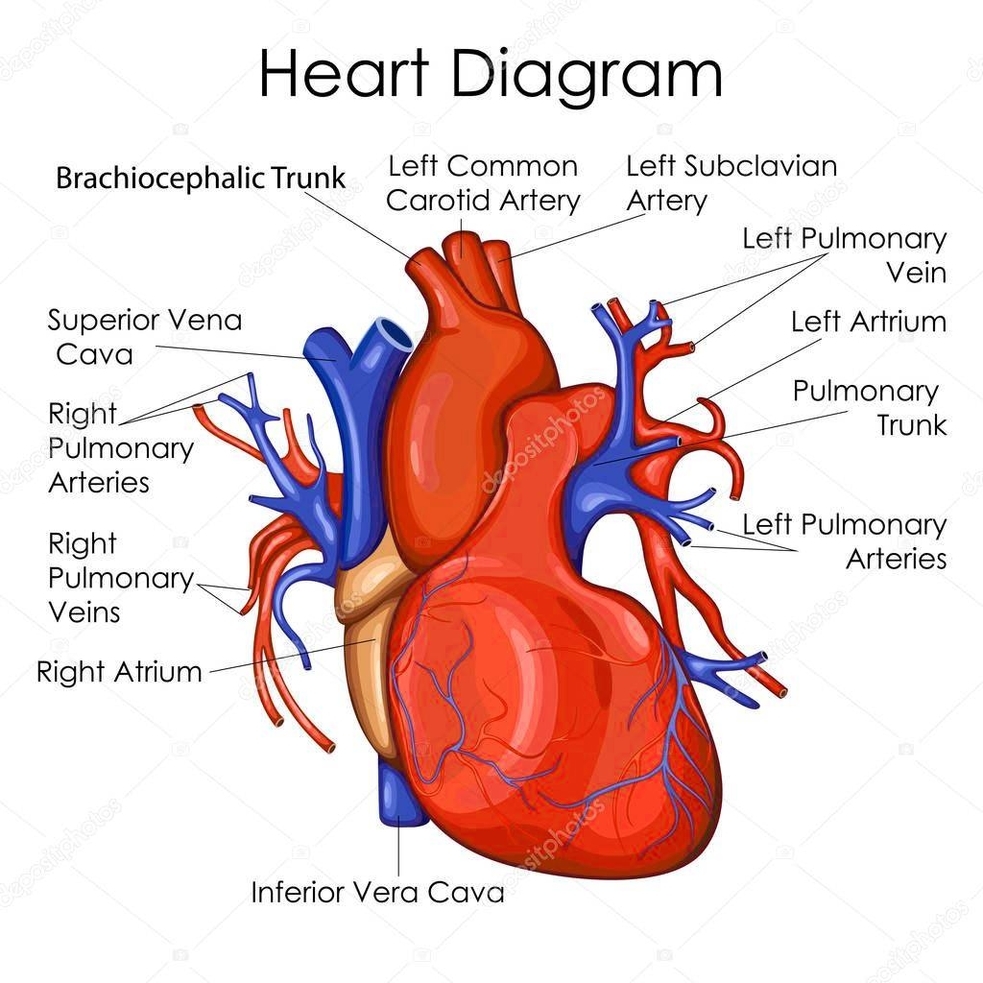
Biology Diagrams Sample Of: Biology diagrams include detailed illustrations of cellular structures, organ systems, and biological processes. Examples include diagrams of the human heart, plant cells, and the process of photosynthesis, aiding in the understanding of complex biological concepts.

Biology Diagrams Sample Of

Womens Size Chart

Womens Size Chart: A women’s size chart provides a standardized guide for selecting clothing sizes based on body measurements, including bust, waist, and hip dimensions. Sizes may be listed numerically (e.g., 2, 4, 6) or alphabetically (XS, S, M, L, XL), and may also offer specialized categories like petite, tall, or plus sizes. These charts are crucial in ensuring proper fit across various brands and styles, especially in an era where sizing can differ significantly by manufacturer or country.

Women's Size Chart