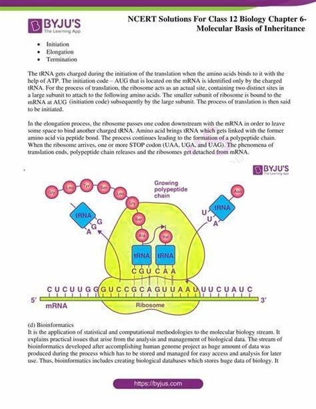
Human Cell with Detailed Labels

Human Cell with Detailed Labels: A detailed diagram of a human cell includes organelles like the nucleus, nucleolus, ribosomes, mitochondria, endoplasmic reticulum, Golgi apparatus, lysosomes, peroxisomes, and centrosomes, along with the plasma membrane and cytoskeleton. Each component is labeled to reflect its function, such as energy production, protein synthesis, or genetic storage. These labels and structures offer insight into how cells operate as the fundamental units of life.

Human C

The Layers of Human Skin

The Layers of Human Skin: The human skin is composed of three primary layers—epidermis, dermis, and hypodermis—each serving unique and vital functions. The outermost layer, the epidermis, provides a waterproof barrier and is responsible for our skin tone. It continuously sheds dead skin cells and regenerates itself. Beneath that lies the dermis, a thicker layer housing nerve endings, hair follicles, sweat glands, and connective tissues that give the skin its elasticity and strength. The innermost layer, the hypodermis (or subcutaneous tissue), consists mainly of fat and connective tissues that insulate the body and act as a cushion to protect internal organs. Understanding these layers helps explain how skin functions as a protective organ and how injuries or diseases impact different levels.

Biological Diagram Online

Sperm Diagram with Labels

Sperm Diagram with Labels: A sperm diagram with labels illustrates the structure of a mature sperm cell, highlighting key components such as the head, midpiece, and tail (flagellum). The head contains the nucleus, which carries the father’s genetic material, and is capped by the acrosome, a specialized organelle containing enzymes that help the sperm penetrate the egg. The midpiece houses mitochondria that supply the energy required for motility. The tail propels the sperm forward using whip-like movements. This diagram is fundamental in understanding male reproductive biology and the journey of fertilization.

Biological Diagrams Visual

Human Reproduction Diagram

Human Reproduction Diagram: A human reproduction diagram displays the male and female reproductive systems, labeled with structures such as the testes, vas deferens, penis, ovaries, fallopian tubes, uterus, and vagina. Additional diagrams may show fertilization, embryo development, and menstrual cycle phases. These illustrations help explain the complex biological process of reproduction, from gamete formation to childbirth, and are fundamental in health education and biology curricula.

Books Th

Eye Anatomy Diagram

Eye Anatomy. Complete Physiology of Eye is described below in the given paragraph: The eye is rather like a living Camera. Each eye is a liquid-filled ball 2.5 cm in diameter. At the front of the eye is a clear, round window called the cornea.

The colored part of the eye. The iris is partly responsible for regulating the amount of light permitted to enter the eye. Lens (also called crystalline lens). The transparent structure inside the eye that focuses light rays onto the retina.

The opening in the middle of the iris through which light passes to the back of the eye. Retina. The light-sensitive nerve layer that lines the inside of the back of the eye. The retina senses light and creates impulses that are sent through the optic nerve to the brain.

Eye Anatomy Diagram

Skin Structure

Here we will discuss the structure of human skin, … and keeps water in. It is composed of layers of flat cells called “squamous” cells.

The Individual Layers of Skin and Their Functions. 1 The Epidermis. The epidermis is the outermost layer of the three layers of skin. Its thickness depends on where it is located on the body. For … 2 The Dermis. 3 The Subcutaneous Tissue.

The skin and its accessory structures make up the integumentary system, which provides the body with overall protection. The skin is made of multiple layers of cells and tissues, which are held to underlying structures by connective tissue (Figure 1). The deeper layer of skin is well vascularized (has numerous blood vessels).

Skin Structure

Brain Structure and Function

The Structure And Function Of The Human Brain. The brain structure is composed of three main parts: the forebrain, midbrain and hindbrain, each with multiple parts. The Cerebrum: Also known as the cerebral cortex, the cerebrum is the largest part of the human brain, and it is associated with higher brain function such as thought and action.

The human brain controls nearly every aspect of the human body ranging from physiological functions to cognitive abilities. It functions by receiving and sending signals via neurons to different parts of the body.T he human brain, just like most other mammals, has the same basic structure, but it is better developed than any other mammalian brain.

Brain Structure & Function offers free color in print and online for all its papers! Brain Structure & Function publishes research that provides insight into brain structure−function relationships.

Brain Structure and Function

Cerebrum Brain Diagram

The cerebrum, originally functioning as part of the olfactory lobes, is involved with the more complex functions of the human brain. In…. The cerebral hemispheres consist of an inner core of myelinated nerve fibres, the white matter, and an outer cortex of gray matter.

The cerebrum is made up of the two cerebral hemispheres and their cortices, (the outer layers of grey matter), and the underlying regions of white matter. Its subcortical structures include the hippocampus, basal ganglia and olfactory bulb.

Hindbrain functions: The three regions of the hindbrain coordinates all processes necessary for survival. These induce breathing, heartbeat, sleep, wakefulness and motor learning. The cerebrum is the largest part of the brain. It consists of the cerebral cortex and other subcortical structures.

Cerebrum Brain Diagram

Worm Anatomy Diagram

Worm Anatomy A worms body is made up of many segments called ‘annuli’. The length of a worms body has muscles which contract and relax which enables the worm to move along a surface. The ‘annuli’ are covered in tiny hairs called ‘setae’ which help the worms movement.

Not necessarily in that order:) The worm has a pointed head and a slightly rounded body. The rings on its body are called segments. Earthworms have no protruding body parts making the worm very contour which enables them to pass through the soil with ease and to also squeeze in between tiny cracks.

Place earthworm in the dissecting tray & rinse off the excess preservative. Identify the dorsal side, which is the worm’s rounded top, and the ventral side, which is its flattened bottom. Turn the worm ventral side up, as shown in the earthworm anatomy diagram below. 3.

Worm Anatomy Diagram

Brain Parts Functions

Parts of the Brain and Their Functions. Cerebrum. The cerebrum is the largest portion of the brain, and contains tools which are responsible for most of the brain’s function. It is divided into four sections: the temporal lobe, the occipital lobe, parietal lobe and frontal lobe.

The outer parts control the more emotional and thought based functions. While the inner and lower parts of the brain control simpler and more basic functions like breathing and blood pressure. Now we will take a trip through the different human brain parts, exploring their function.

Although each structure has a distinct function, they work together to control all functions of the body. The brain is made up of 3 essential parts: Cerebrum, Cerebellum, and Brainstem. The cerebrum is the largest part of the human brain. It has a rough surface (cerebral cortex) with gyri and sulci.

Brain Parts Functions