See the below image for the Biology cell free vector graphic on pixabay0 diagram.
Charts, Graphs and Diagrams
See the below image for the Biology cell free vector graphic on pixabay0 diagram.
See the below image for the Biology cell structure diagram.
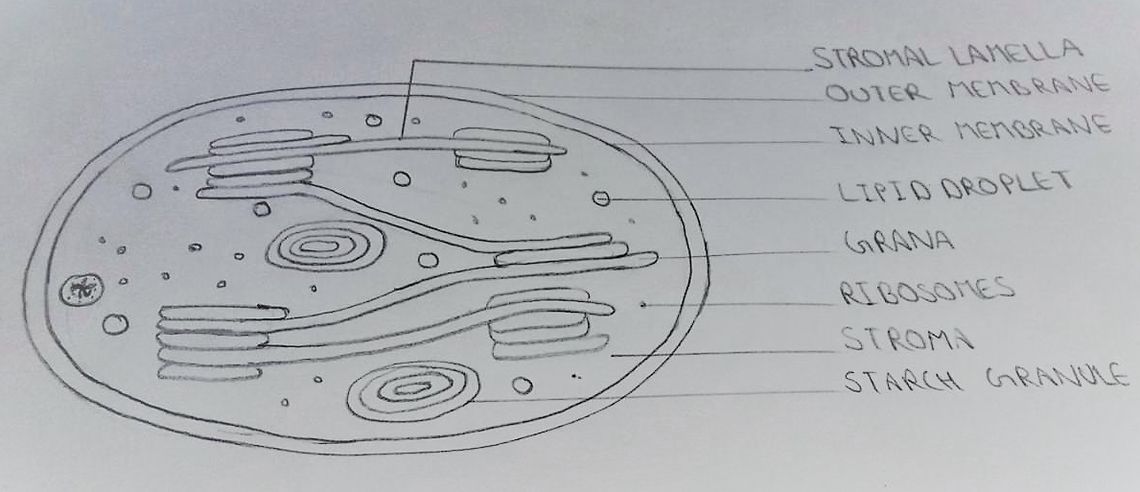
See the below image for the How to draws in biology diagram. Biological drawings must be done on unlined sheets or plane sheets, for more authenticity. This principle should be followed particularly when you are drawing a diagram for your biology lab copy or record. But don’t worry about your board exam you get lined copy and you have to draw on that lined sheet. 6.
Drawing Materials: All drawings should be done with a sharp pencil line on white, unlined paper. Diagrams in pen are unacceptable because they cannot be corrected. Positioning: Center drawing on the page.
Biological drawings should be conspicuous for easier comprehensibility- Avoid crowding components of the diagram. A biological drawing must bear all relevant parts that are conspicuous enough to the human eye. It should also be large enough to current the whole complicated background details of the diagram to the observer.
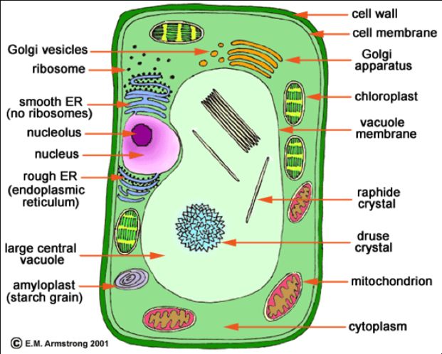
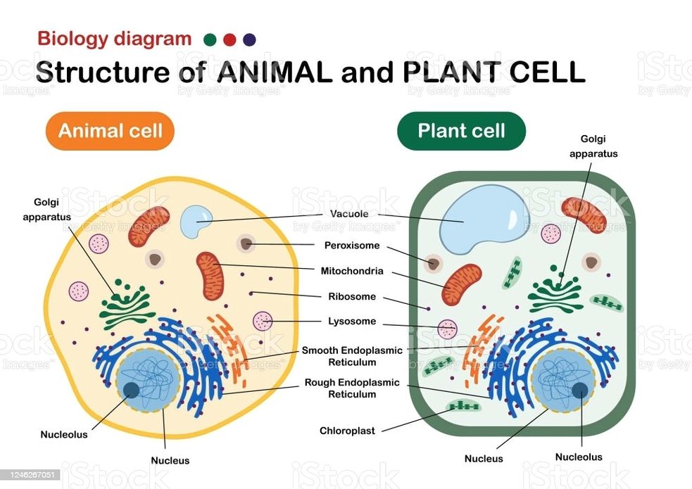
See the below image for the Biology show structure of animal and plant cell diagram. Plant and Animal Cell Structures 1 They are single membrane-bound, fluid-filled structures. 2 They are found in both plants and animal cells. Plant cells have large, central vacuole. … 3 Tonoplast is the membrane of these vacuoles. 4 They maintain turgor pressure.
Structure of Animal and Plant Cells 1 Have a nucleus 1. Plant cells have a cellulose cell wall 2 Have a cytoplasm 2. Plant cells have a vacuole containing cell sap 3 Have a cell membrane 3. Plant cells have chloroplast 4 Contain mitochondria 4. Many plant cells have a box-like shape whilst animal cell shape varies 5 Contain ribosomes
Generalized Structure of a Plant Cell Diagram. As you can see in the above labeled plant cell diagram under light microscope, there are 13 parts namely, Cell membrane. Cytoplasm. Ribosomes. Nucleus.
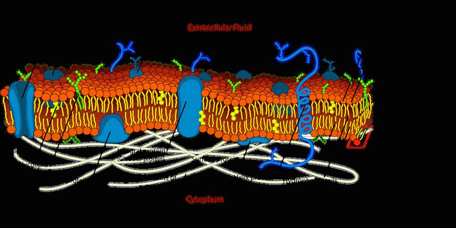
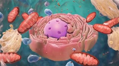
See the below image for the Cell biology cell structure and functions diagram. The cell structure comprises individual components with specific functions essential to carry out life’s processes. These components include- cell wall, cell membrane, cytoplasm, nucleus, and cell organelles. Read on to explore more insights on cell structure and function.
The cell function is to keep all of the functions of the body performing as intended. This includes keeping toxins out of the body, help to break down waste, make nutrients and act as barriers within organelles.
The nucleus protects the DNA and is an integral component of a plant’s cell structure. Cells are composed of various cell organelles that perform certain specific functions to carry out life’s processes.
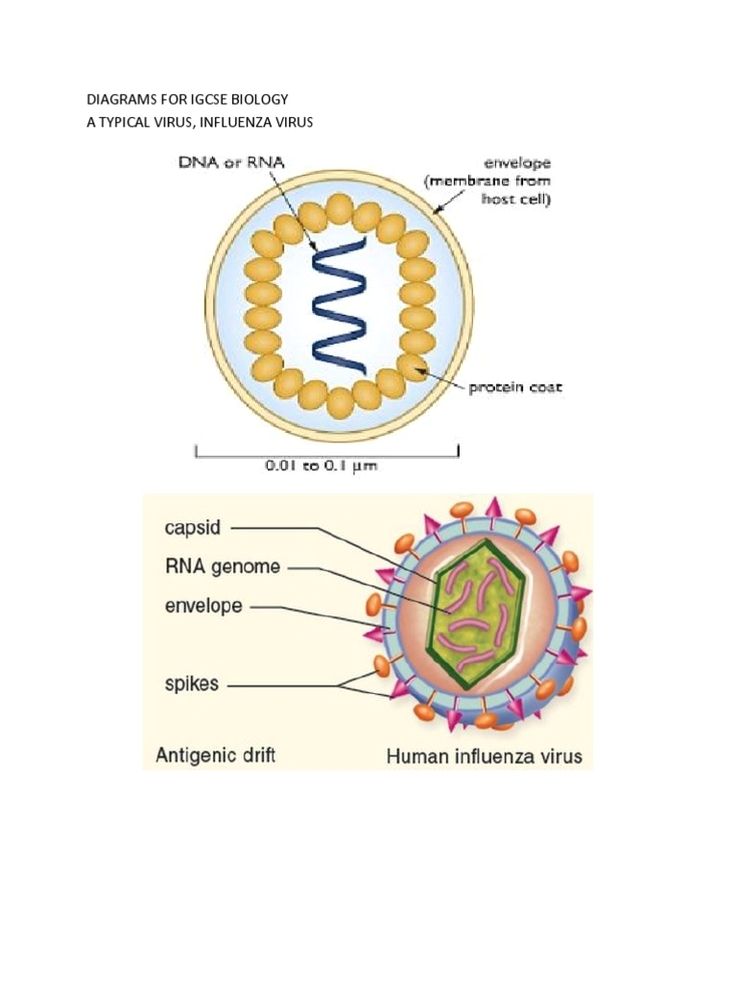
See the below image for the s for igcse biology diagram. IGCSE Biology Past Papers. Complete IGCSE Biology Past Papers. With an emphasis on human biology, the Cambridge IGCSE Biology syllabus helps learners to understand the technological world in which they live, and take an informed interest in science and scientific developments.
Cambridge IGCSE Biology (0610) Cambridge IGCSE Biology helps learners to understand the biological world in which they live and take an informed interest in science and scientific developments.
CIE IGCSE Biology Revision. Papers 1-6. Topic 1: Characteristics and Classification of Living Organisms. Topic 2: Organisation of the Organism. Topic 3: Movement in and out of Cells. Topic 4: Biological Molecules. Topic 5: Enzymes. Topic 6: Plant Nutrition. Topic 7: Human Nutrition. Topic 8: Transport in Plants. Topic 9: